
Listening to the pulse
Structuring the imaging question in pulsatile tinnitus
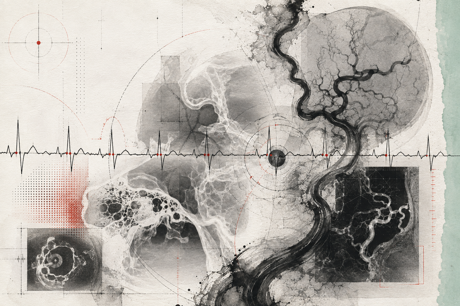
Which anatomical circuit could plausibly create the perceived waveform?
Six ingredients,
one interpretation.
Four movements.
Characterise
Clarify timing, synchrony, laterality and associated neurological or visual features.
Localise
Use clinical clues to organise arterial, venous and non-vascular possibilities.
Select
Match the imaging pathway to the unresolved anatomical question.
Escalate
Recognise when non-invasive findings remain discordant with clinical concern.
Synchrony.
The temporal relationship between the perceived sound and the pulse helps organise the question before imaging attempts to answer it.
PULSE / CIRCUIT / CAUSEHold both sides
in view.
Expert synthesis is rarely a choice between simple opposites. The work is to understand how each tension changes confidence.
Make the reasoning visible.
Describe the symptom precisely, map plausible arterial, venous and non-vascular circuits, and make the reason for each imaging step explicit.
“First listen to the rhythm. Then decide where to look.”Clinical Recipes / Editorial reflection
Three deeper
readings.
RHYTHM → CIRCUIT → TARGET
Treat the symptom as a waveform
Ask the patient to describe timing, laterality, constancy and modulation. Pulse synchrony is not a diagnosis, but it changes the map of plausible generators and gives the imaging conversation a useful first coordinate.
Draw competing circuits
Arterial inflow, venous outflow and non-vascular transmission can produce similar language at the bedside. Keep several circuits visible long enough to test each against examination and non-invasive imaging.
Escalate for a named uncertainty
Further imaging is most useful when it answers an explicit residual question. The reason to proceed should be written before the next study—not invented after an incidental finding appears.
Questions for
colleagues.
- 01
Is the sound truly pulse-synchronous?
- 02
What manoeuvre changes it, and what circuit does that suggest?
- 03
Which unresolved question justifies the next modality?
The ear reports a rhythm; the clinician must decide which circulation could plausibly conduct it.
Pulsatile tinnitus has diverse causes; evaluation and imaging selection require specialist judgement.