
Decoding the nidus
Reading AVM angioarchitecture as a connected system
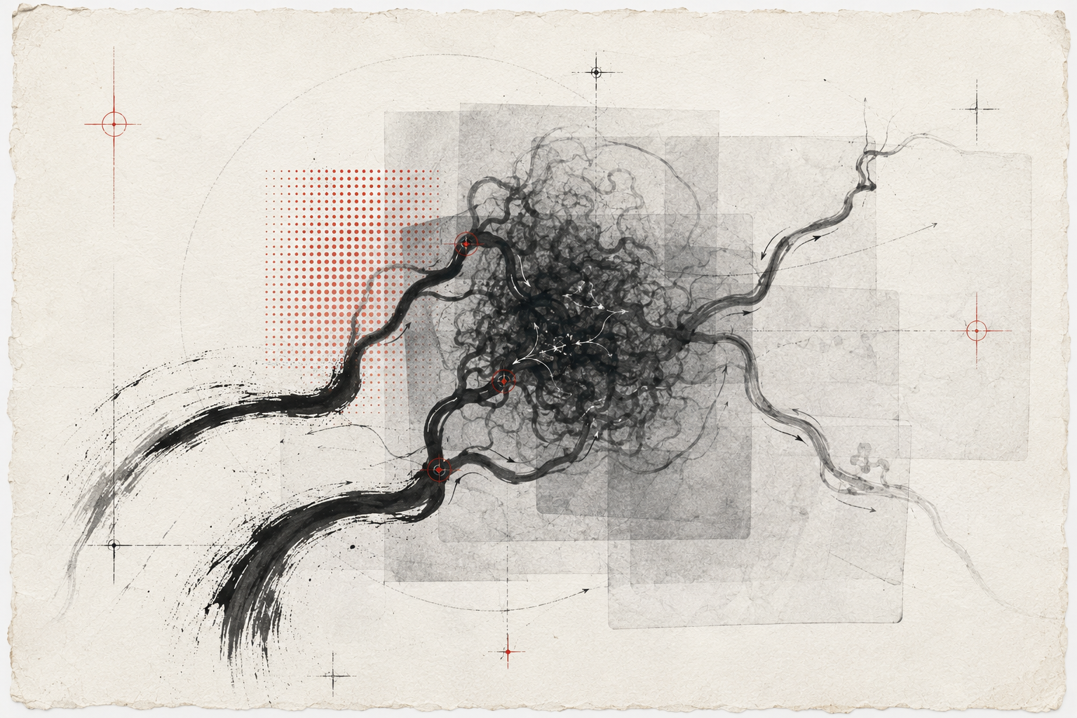
Where does the network concentrate vulnerability or procedural consequence?
Six ingredients,
one interpretation.
Four movements.
Map
Name the components before compressing them into a grade or label.
Sequence
Read arterial arrival, nidal transit and venous egress over time.
Relate
Connect architecture to presentation, haemodynamics and surrounding eloquence.
Revisit
Expect architecture to evolve spontaneously or after partial treatment.
Sequence.
An AVM is not only a collection of vessels. Time-resolved filling reveals how feeders, nidus and venous drainage behave as a connected haemodynamic system.
INFLOW / NIDUS / OUTFLOWHold both sides
in view.
Expert synthesis is rarely a choice between simple opposites. The work is to understand how each tension changes confidence.
Make the reasoning visible.
Present the architecture in order: inflow, nidal transit, outflow and associated features. Preserve what remains uncertain or incompletely visualised.
“The architecture becomes legible when anatomy is allowed to move through time.”Clinical Recipes / Editorial reflection
Three deeper
readings.
INFLOW → NIDUS → OUTFLOW
Keep the network intact
A list of feeders and veins is necessary but insufficient. The lesion becomes legible when components are read as a connected system with preferential routes, compartments and zones of haemodynamic concentration.
Let time disclose architecture
Early arrival, intranidal transit and venous egress separate structures that overlap on a static projection. Temporal order helps distinguish supply from passage and passage from drainage.
Name what may change
Associated aneurysms, venous restriction and compartmental filling can evolve with natural history or partial treatment. A useful description preserves a baseline that future teams can truly compare.
Questions for
colleagues.
- 01
Where is flow concentrated rather than merely present?
- 02
Which feature is visible only because of temporal separation?
- 03
What architecture must be preserved in the longitudinal record?
The nidus is not an object on an image; it is a vascular relationship unfolding in time.
Angioarchitecture is multidimensional and may change over time; management requires multidisciplinary expertise.