
Making time visible
A disciplined longitudinal imaging comparison
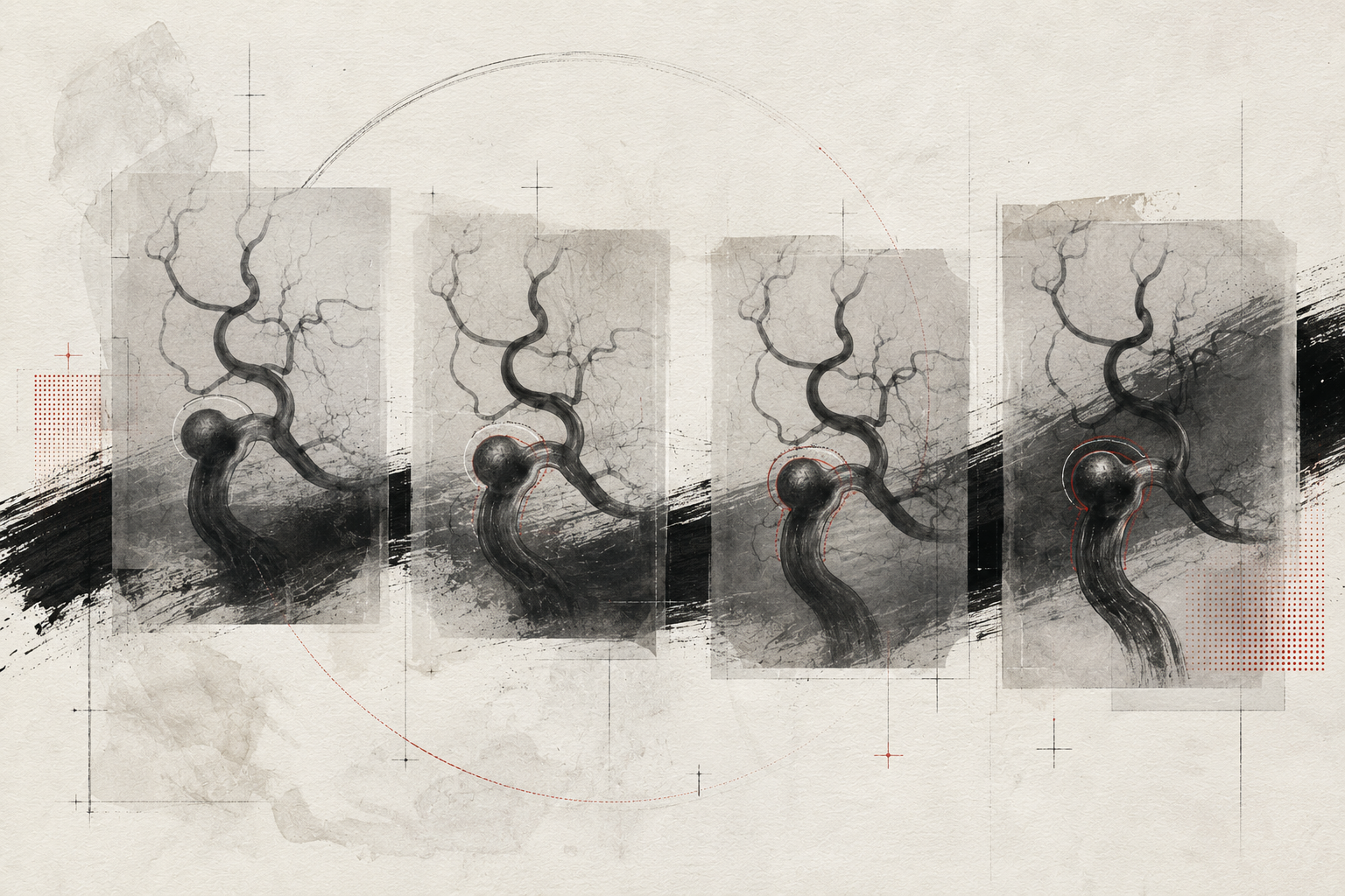
Is the lesion changing, or did the way we look at it change?
Six ingredients,
one interpretation.
Four movements.
Anchor
Choose the earliest technically reliable study as the reference.
Align
Compare like with like and disclose changes in technique.
Detect
Look beyond diameter for new irregularity, thrombus or wall signal.
Record
Document both change and confidence in the observation.
Registration.
Before declaring biological change, make the studies comparable. Plane, reconstruction, contrast timing and measurement method can manufacture an apparent interval.
BASELINE / INTERVAL / CONFIDENCEHold both sides
in view.
Expert synthesis is rarely a choice between simple opposites. The work is to understand how each tension changes confidence.
Make the reasoning visible.
Pair images, disclose technical differences and describe both the observed change and confidence in its reality. Let the chronology remain visible.
“Time clarifies only when the images truly meet.”Clinical Recipes / Editorial reflection
Three deeper
readings.
BASELINE → REGISTRATION → CHANGE
Choose a baseline that can carry time
The first available study is not always the best reference. Select the earliest technically reliable examination and document why its acquisition, planes and reconstructions can support comparison.
Make the studies meet
Align equivalent projections and source planes before measuring. Contrast timing, slice thickness and reconstruction algorithms can create persuasive but artificial differences when viewed side by side.
Separate observation from confidence
Describe diameter, contour, thrombus, wall signal and adjacent anatomy, then state how certain the interval change is. A small change with high technical uncertainty is a different finding from reproducible morphological evolution.
Questions for
colleagues.
- 01
Are these studies technically comparable?
- 02
Which descriptor changed beyond diameter alone?
- 03
How confident are we that the interval is biological?
Longitudinal imaging is not two pictures; it is a carefully constructed relationship between them.
Apparent interval change can reflect acquisition, reconstruction or measurement differences.